Screening
High content screening (HCS) is used to identify compounds such as small molecules that result in a desired change to the phenotype of a cell, micro-tissue or model organism. This is typically performed on cells in culture and may, for example, measure change in expression, redistribution or translocation of proteins or change in morphology, functional performance or viability of each cell.
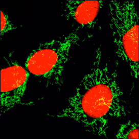
Cells are exposed to the substance(s) and thereafter the morphology and components of these cells are interrogated. Commonly this involves labelling proteins with fluorescent tags and changes in the cell phenotype are measured using automated imaging or alternatively, for non-adherent cells, with (high throughput) flow cytometry. Imaging can detect subcellular changes while flow cytometry can measure many fluorescent parameters simultaneously.
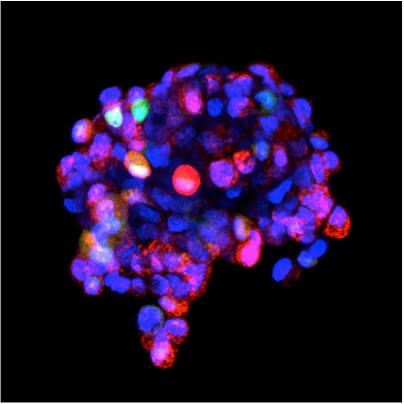
Image-based screening is essentially automated fluorescence microscopy performed on large numbers of samples, commonly of adherent cells in wells of micro plates.
Use of a fluorescent DNA probe allows counterstaining and therefore location of individual cells, segmentation of their nuclei and total cell enumeration. A cell-permeant counterstain permits choice of either live- or fixed-cell endpoint assay and simple transfer between the two.
A fluorescent viability probe can be utilized in live-cell assays to help establish levels of cellular toxicity, apoptosis induction and cell health.
Non-adherent cells or microtissues or motile organisms may require immobilisation to permit reliable and high-performance imaging of structures and changes.
Related BioStatus Products for Image-Based Screening
DRAQ5™ in Image-Based Screening
Far-red DNA counterstain, nucl:cyto segmentation in live- / fixed-end point assays.
DRAQ7™ in Image-Based Screening
Reliable identification of apoptotic/dead cells in real-time.
CyTRAK Orange™ in Image-Based Screening
Single-channel nucl:cyto segmentation in live- / fixed-end point assays.
CyGEL™ in Image-Based Screening
Convenient immobilization of live non-adherent / motile objects.
CyGEL Sustain™ in Image-Based Screening
Culture medium-specific immobilization of live non-adherent / motile objects.
High-throughput flow cytometry permits rapid sequential analysis of cell phenotypes in large sets of treated samples and controls for non-adherent cell types, blood and bone marrow.
Cell-permeant fluorescent DNA dyes can be used to discriminate nucleated from anucleated cells reducing the need for time-consuming and potentially damaging RBC lysis reagents.
Stoichiometric DNA labeling can provide information on cell cycle status, ploidy and proliferation (S-phase fraction) without any permeabilization or RNase treatment.
A fluorescent viability probe can be utilized to evaluate sample integrity, cell death or apoptosis in live cell assays.
Related BioStatus Products for High Throughput Flow Cytometry
DRAQ5™ in High Throughput Flow Cytometry
No-lyse, no-wash analysis of samples & Direct DNA content analysis.
DRAQ7™ in High Throughput Flow Cytometry
Simple dead cell exclusion & clear identification of apoptotic/dead cells.
CyTRAK Orange™ in High Throughput Flow Cytometry
No-lyse, no-wash analysis of nucleated cell samples.
In-Cell Western™ assays measure the change of protein expression under different conditions for a total cell population in a well. After treatment/exposure the cells are fixed and permeabilised to allow access of a fluorescently-tagged antibody to label the protein of interest.
A fluorescing stoichiometric DNA probe is normally applied to the cells in each well. The resulting signal is directly proportional to the number of cells and can be used to normalise the protein expression appropriately.
Related BioStatus Products for In-Cell Westerns™
DRAQ5™ in In-Cell Westerns™
Convenient and predictable normalisation of protein expression versus cell numbers.