Imaging
Fluorescence Microscopy
Fluorescence microscopy uses fluorescence instead of, or in addition to, transmission and diffraction to study properties of cells, tissues and small organisms by measuring the signal from a fluorescent probe or a fluorescently-tagged antibody, for example.
This can be done by epifluorescence microscope (the most common and simplest of designs delivering high sensitivity but low spatial resolution), confocal microscope (for higher spatial point resolution, typically based on a scanning laser or Nipkow spinning disc to permit 3-D sectioning of a sample in the z- plane), light sheet microscopy or super-resolution techniques such as STED or 3D- SIM (aka “OMX”).
All of these methods rely upon simple excitation and emission characteristics of fluorescent molecules. These techniques are used for fixed or living cells, tissues and organisms.
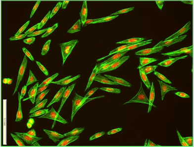
Immunofluorescence involves microscopical analysis of cells and paraffin-embedded or frozen tissue sections. Samples are fixed (usually with formaldehyde) to preserve the phenotype and permeabilized to allow fluorescently-labelled antibodies access to the internal antigens.
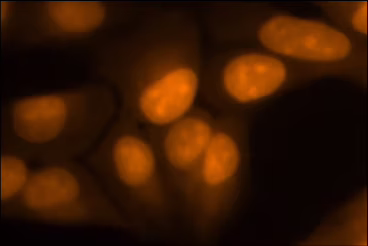
Use of a fluorescent DNA probe allows counterstaining and therefore location of individual cells, segmentation of their nuclei and total cell enumeration.
Non-adherent cells, micro-tissues or motile organisms may require immobilisation to permit reliable and high-performance imaging of structures and changes.
Related BioStatus Products for Immunofluorescence Microscopy
DRAQ5™ in Immunofluorescence Microscopy
Far-red fluorescing DNA counterstain; GFP/FITC compatible, no UV needed.
CyTRAK Orange™ in Immunofluorescence Microscopy
Orange fluorescing DNA counterstain; GFP/FITC compatible, no UV needed.
CyGEL™ in Immunofluorescence Microscopy
Convenient immobilization of non‐adherent or fragile objects
In some cases it is essential to keep cells or tissue alive and intact while imaging as the cell-processing used in immunofluorescence techniques destroys the feature of interest, causes its redistribution or loss on washing.
It may also be convenient to avoid such cell processing when the feature of interest is tagged with a fluorescent protein (e.g. GFP). Cell-permeant fluorescent DNA dyes can be used as a counterstain.
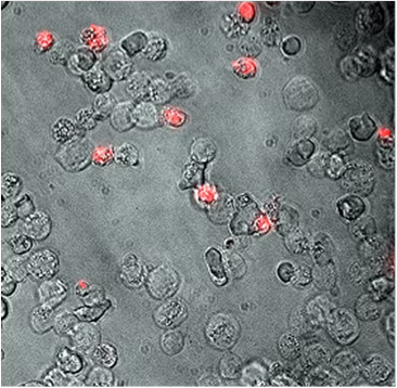
A fluorescent viability probe can be utilized to evaluate sample integrity, cell death or apoptosis in live-endpoint assays.
Live non-adherent cells, micro-tissues or motile organisms may require immobilisation to permit reliable and high-performance imaging of structures and changes.
Related BioStatus Products for Live-Endpoint Cell/Tissue Imaging
DRAQ5™ in Live-Endpoint Cell/Tissue Imaging
Far-red fluorescing live-cell permeant DNA counterstain.
DRAQ7™ in Live-Endpoint Cell/Tissue Imaging
Clear identification of damaged/apoptotic/dead cells.
CyTRAK Orange™ in Live-Endpoint Cell/Tissue Imaging
Orange fluorescing live-cell permeant DNA counterstain.
CyGEL™ in Live-Endpoint Cell/Tissue Imaging
Convenient immobilization of live non‐adherent or fragile objects.
Organisms such as zebrafish and drosophila embryos, C. elegans and parasites are useful subjects for imaging but their high motility means that they require immobilization to gain useful information.
The onward growth or orthogonal analysis after imaging requires gentle and easy recovery from the immobilizing agent.
Related BioStatus Products for Live Organism Imaging
CyGEL™ in Live Organism Imaging
Convenient thermo-reversible immobilization of live motile organisms.
CyGEL Sustain™ in Live Organism Imaging
Culture medium-specific, thermo-reversible immobilization of live motile organisms.