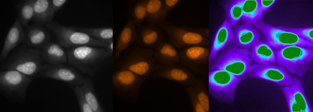
We find that DRAQ5™ is the most robust DNA dye we have ever used.
It stains nuclei much brighter than cytoplasm and allows us to obtain clear images from even the most difficult tissues.
Prof. Melinda K Duncan
University of Delaware
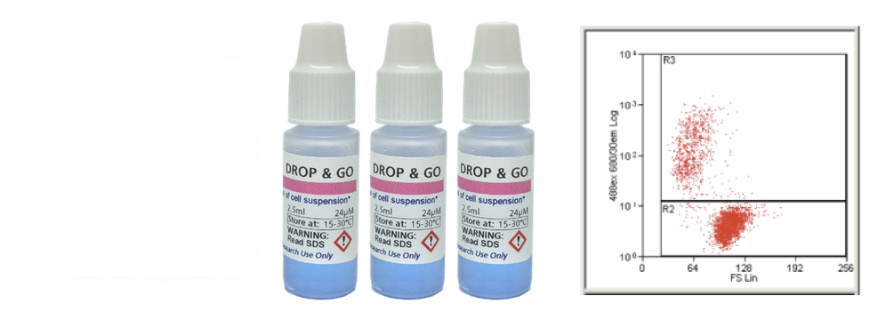
DRAQ7™ has enabled us to do experiments that were not otherwise possible before.
Not only is it a much cleaner signal than other viability dyes, but the fact that it is non-toxic has enabled us to do time courses with the same samples.
Highly recommended!
Dr Gareth Griffiths
Imagen Biotech
We found that CyGEL™ was suitable for complete immobilisation of procyclic stage L. major and T. brucei for 90 minutes without affecting cell viability.
This has enabled us to analyse a GFP-tagged form of the L. major membrane-associated protein, HASPB, using live confocal imaging and FRAP.
Dr Helen Price
DRAQ7™ is a far-red fluorescent DNA dye that ONLY stains the nuclei in DEAD and permeabilized cells. DRAQ7™ is an ideal viability dye to report cell health over extended time periods.